



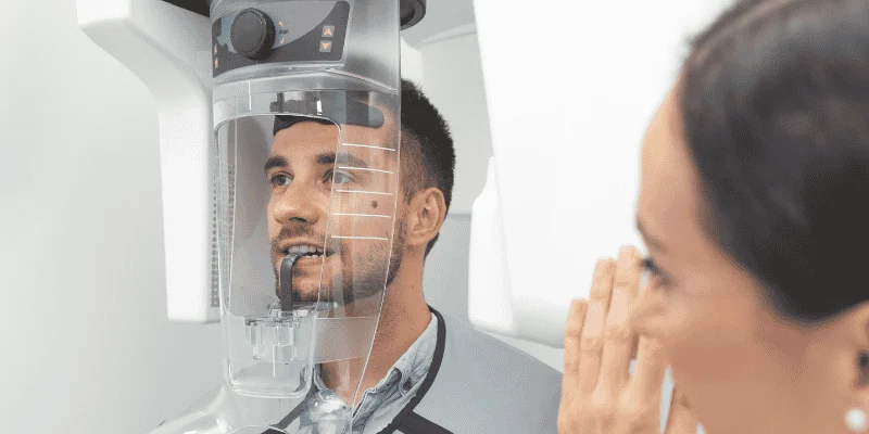
You may think regular dental technology, such as X-ray, shows everything going on in your mouth. It gives a flat image of your teeth and jaw. But it cannot show depth, hidden angles, or detailed views of nerves and bone.
A 3D cone beam scan lets your dentist see your teeth, jaw, nerves, and bone in three dimensions, which helps them find problems that a flat X-ray can miss.
This type of scan rotates around your head and builds a full 3D image.
You get a clearer view of impacted teeth, small infections, bone loss, and the exact position of nerves.
When your dentist plans implants, treats jaw pain, or looks for the cause of ongoing discomfort, that extra detail matters. With a 3D view, you get more precise planning and fewer surprises during treatment.
Key Takeaways
- A 3D scan shows detailed views of teeth, bone, and nerves that flat X-rays cannot show.
- Dentists use 3D imaging to plan complex treatments with greater accuracy.
- The added detail helps detect hidden problems and improve treatment decisions.
Key Differences Between 3D Cone Beam CT and Regular Dental X-Rays
When your dentist chooses between a 3D cone beam CT and a regular dental X-ray, they focus on depth, detail, and purpose. Each tool captures different information about your teeth, roots, bone, and nearby structures.
How CBCT Imaging Works
A CBCT scan uses cone beam computed tomography to capture your teeth and jaws in three dimensions. The CBCT scanner rotates around your head and collects many images in one sweep.
The system rebuilds these images into a 3D dataset. Your dentist can view thin slices from different angles, including front to back and side to side. This process is called CBCT imaging.
Because it shows depth, a dental cone beam CT helps your dentist see exact root positions, bone width, nerve canals, and sinus spaces. It reduces the problem of structures overlapping each other.
Dentists often rely on 3D scans for implants, surgery, and complex root canal cases. The main advantage is spatial accuracy.
How Traditional Dental X-Rays Work
Traditional dental X-rays capture flat, two‑dimensional images. A small sensor or film sits inside your mouth while the machine sends a focused beam through your teeth.
The result is a single projection. It compresses 3D anatomy into one image.
Because of this, structures can overlap. For example, a root may hide behind another root, or bone levels may look slightly different depending on the angle.
Still, regular dental X-rays remain essential. They use low radiation and work well for routine exams, cavities, and basic bone checks; they provide enough information.
Comparing Scan Time, Image Detail, and Field of View
You will notice differences in scan time, image detail, and field of view between these tools.
A panoramic X-ray or periapical image usually takes seconds. A cone beam CT scan also captures data quickly, but reviewing the full 3D dataset takes more time because your dentist must examine many slices.
Image detail also differs:
| Feature | Regular Dental X-Rays | 3D Cone Beam CT |
| Image type | Flat 2D image | 3D volume |
| Overlap | Structures may overlap | Minimal overlap |
| Depth | Estimated | Clearly visible |
| Field of view | Small or full arch (panoramic) | Adjustable, small to large |
A larger field of view lets your dentist see the full jaw, airway, or both arches. A smaller one targets a single tooth. This flexibility makes dental cone beam scans useful when location and distance truly matter.
Types of Dental X-Rays: Periapical, Panoramic, and More
Your dentist uses different types of traditional dental X-rays based on the question they need to answer.
Periapical X-rays show one or two teeth from crown to root tip. Dentists use them to check infections, root shape, and bone around the apex.
Panoramic X-rays capture the entire upper and lower jaws in one wide image. They help screen for impacted teeth, jaw issues, and general bone patterns.
Other images, like bitewings, focus on cavity detection between teeth.
These 2D tools remain the first step in most visits. When they cannot clearly show depth or exact position, your dentist may recommend a cone beam ct to gain the extra detail needed for safe and accurate treatment planning.
What CBCT Reveals That 2D X-Rays Miss
A CBCT scan shows depth, position, and structure in ways flat dental x-rays cannot. You see canals, nerves, bone shape, and small defects in three dimensions instead of guessing from a single projection.

Hidden Root Canals and Tooth Fractures
When you look at periapical x-rays, you see one flat image. Roots overlap, and small details can hide behind each other.
With a cone beam CT scan, you scroll through thin slices of the tooth from different angles. This helps you spot extra root canals that a standard periapical x-ray may miss. Missed canals often explain why symptoms continue after treatment.
CBCT imaging also helps you check for vertical root fractures. These cracks can be hard to see on dental x-rays because the fracture line may run in the same direction as the X-ray beam.
A dental cone beam CT lets you view the root from the side, front, and top. That clear view helps you decide if retreatment, surgery, or extraction makes sense.
For complex endodontic cases, dentists often rely on 3D imaging because it shows canal shape and curvature more clearly than 2D films.
Many clinicians use CBCT when standard imaging leaves important questions unanswered.
Precise Nerve and Sinus Mapping
In the lower jaw, the inferior alveolar nerve runs close to tooth roots. On a regular panoramic or periapical x-ray, you can only estimate how near that nerve sits to a root or implant site.
A CBCT scan shows the nerve canal in cross-section. You measure the exact distance between the root tip and the nerve. This reduces the risk of numbness after surgery.
In the upper jaw, the maxillary sinus often sits just above molar roots. A cone beam CT scan shows whether roots extend into the sinus or only touch its floor.
This level of detail matters for extractions, implants, and sinus lift planning. A panoramic X-ray may show height, but it does not show width or true spatial position. Dentists often upgrade imaging in these cases.
Identifying Periapical Lesions and Bone Defects
Periapical lesions form at the tip of a tooth root, usually from infection. On periapical x-rays, small lesions can hide behind thick bone or overlap with nearby structures.
CBCT imaging shows the area in thin slices. You see the true size, shape, and spread of the lesion. This helps you tell the difference between a small infection and a larger cyst-like defect.
You also detect bone defects on the cheek or tongue side of the root. These defects may not appear on 2D dental imaging because the bone overlaps in a flat image.
A dental cone beam scan reduces superimposition, which means fewer hidden areas. Dentists use this added detail when symptoms do not match what periapical x-rays show. In these situations, a CBCT changes treatment planning because it answers spatial questions that 2D images cannot.
Accurate Bone Density and Structure
When you plan an implant, you need to know more than bone height. You need bone width, contour, and density.
A cone beam CT scan shows the ridge in cross-section. You measure how thick the bone is from front to back and from cheek to tongue. This prevents placing an implant where bone is too thin.
CBCT imaging also gives clues about bone density. While it does not replace medical CT for exact density numbers, it helps you judge whether bone looks dense and stable or soft and porous.
Regular dental x-rays cannot show this three-dimensional structure. A 3D scan captures a full volume of the jaw, which allows careful review of anatomy before surgery.
Modern systems provide detailed views with lower radiation than medical CT.
Applications of CBCT in Modern Dental Care
A 3D cone beam scan gives you detailed views of bone, teeth, nerves, and soft tissue. Dentists use it to plan procedures with more control and fewer surprises.
Advanced Dental Implant Planning
When you need dental implants, your dentist must know the exact height, width, and density of your jawbone. A regular X-ray flattens these structures. A CBCT scan shows your bone in three dimensions, which improves dental implant planning.
With 3D imaging, your dentist can measure the distance to the sinus or the inferior alveolar nerve. This helps avoid nerve injury and sinus problems.
Many practices rely on 3D CBCT imaging for dental implant treatment planning because it offers more precise measurements than 2D films.
The scan also helps create a surgical guide. This guide directs the angle and depth of the implant during surgery. An oral and maxillofacial radiologist may review the scan to confirm safe implant placement before your procedure.
Managing Impacted and Wisdom Teeth

Impacted teeth often sit at odd angles or press against nearby roots. A standard X-ray may not show how close they are to a nerve.
A CBCT scan shows the exact position of impacted teeth and wisdom teeth in relation to the nerve canal.
This matters most in the lower jaw. If a wisdom tooth touches the nerve, removal carries more risk. A 3D scan helps your dentist plan a safer extraction path.
CBCT has become common in oral surgery because it provides detailed 3D views of bone and tooth position.
Many clinicians use it as part of modern digital workflows in dentistry, including cone beam computed tomography in clinical dental care.
Clear imaging supports better treatment planning and fewer surprises during surgery.
Detecting Gum Disease and Jaw Pathology
Advanced gum disease can destroy bone around your teeth. A 2D X-ray may miss early bone defects, especially on the cheek or tongue side of the tooth. CBCT shows bone loss in all directions.
Your dentist can see:
- Vertical bone defects
- Bone craters between teeth
- Areas of bone thinning
- Hidden infections at the root tips
CBCT also helps detect cysts, tumors, and jaw abnormalities. It creates a 3D view of the maxillofacial region, which improves diagnostic accuracy in many dental fields.
When your provider sees the full shape and depth of bone damage, they can design a more specific and focused treatment plan.
Supporting Orthodontics and Maxillofacial Surgery
If you need braces or jaw surgery, your dentist or orthodontist must study how your teeth and jaws align. A CBCT scan shows tooth roots, jaw position, and airway space in one image.
For orthodontics, this helps identify:
- Impacted canines
- Root position before tooth movement
- Bone thickness around teeth
In oral and maxillofacial surgery, CBCT supports planning for jaw correction, trauma repair, and temporomandibular joint evaluation.
Research shows that CBCT improves diagnosis and planning across implant dentistry, orthodontics, and jaw assessment.
When your care team sees your anatomy in 3D, they can plan movements and surgical steps with more control and accuracy.
Radiation Exposure and Safety Considerations
You want clear answers about radiation exposure before you agree to a 3D scan. Dentists measure radiation dose in millisieverts (mSv) and follow strict radiation safety rules to keep your risk low.
Comparing Radiation Dose: CBCT vs. Standard X-Rays
A cone beam CT (CBCT) scan uses more radiation than a single dental X-ray. However, the actual radiation dose depends on the scan size and settings.
A small field CBCT that looks at one area can expose you to a dose similar to a full set of digital dental X-rays. Larger scans that capture the whole jaw increase the mSv level.
Digital intraoral X-rays use very low radiation. Panoramic X-rays fall in the middle. CBCT gives more detail in three dimensions, so it often requires a higher radiation dose than 2D images.
The key point is purpose. Your dentist should order CBCT only when regular X-rays cannot show enough detail.
Understanding MSV, Background Radiation, and Shielding
Dentists measure radiation exposure in millisieverts (mSv). This unit reflects how radiation may affect your body.
To give context, you receive background radiation every day from natural sources like soil, air, and space. Medical and dental imaging adds to that total.
A CBCT scan adds a small fraction of your yearly background radiation. The exact mSv depends on the machine, scan size, and exposure time.
Shielding also plays a role in radiation safety. Some offices use thyroid collars or protective aprons when appropriate. Research continues on added protection methods for sensitive areas during CBCT scans.
Best Practices for Patient Safety
You reduce unnecessary radiation exposure when you ask why a scan is needed. Your dentist should explain how the result will change your treatment plan.
Clinics follow federal and state rules for dental imaging equipment. They must maintain machines, check calibration, and train staff in proper positioning and exposure settings.
The FDA outlines patient-focused guidance on dental cone-beam computed tomography safety.
Dentists also follow the ALARA principle. This means they keep radiation “as low as reasonably achievable.”
They limit the scan area, use the lowest effective settings, and avoid repeat scans unless truly needed. These steps protect you while still giving your dentist the 3D detail that regular X-rays cannot provide.
Cost, Accessibility, and Who Reads Your CBCT Scan
A dental CBCT scan gives detailed 3D images, but it costs more than a regular X-ray and is not used for every visit. You also want to know who reviews your scan and how trained they are to read it.
When CBCT Is Necessary—And When It’s Not
Your dentist orders CBCT imaging when a flat X-ray cannot show enough detail. This often happens before dental implants, complex root canal treatment, impacted teeth removal, or when nerve position matters.
A cone beam scan helps your dentist measure bone width, see hidden canals, and check for cysts or infections that overlap on 2D images. The 3D view reduces guesswork.
CBCT is not a routine screening tool. Dentists weigh the benefit against radiation exposure. You will not need it for simple cavities, basic cleanings, or most routine exams.
Cost varies by region and scan size. In the United States, a CBCT scan in a dental office often ranges from about $200 to $1,000. Insurance may cover part of the fee when the scan supports a specific procedure, such as implant placement.
Availability in Dental Offices
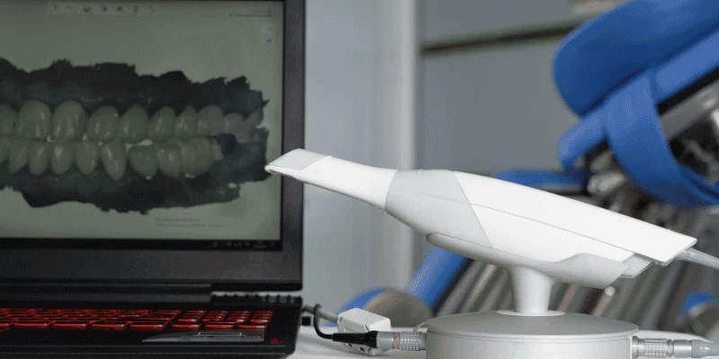
Many specialists now have in-office CBCT systems. Oral surgeons, endodontists, and periodontists often invest in a CBCT scanner because they place implants or treat complex cases.
General dentists may also have a CBCT scanner, especially in larger or group practices. The machine looks similar to a panoramic unit and rotates around your head in one pass. You usually stand or sit still for less than a minute.
If your dentist does not have in-office CBCT, they may refer you to a nearby imaging center. The images are stored digitally and shared with your provider for review and treatment planning.
The Role of the Oral and Maxillofacial Radiologist
Not every CBCT scan is read the same way. Your treating dentist often reviews the scan first because they know your clinical history and treatment goals.
In more complex cases, an oral and maxillofacial radiologist may interpret the images. This specialist completes extra years of training focused on head and neck imaging and radiation safety.
They review the entire scan, not just the area of interest. That includes sinuses, jaw joints, airway spaces, and surrounding bone. Their report can identify findings your dentist may not expect.
Some practices send scans electronically for remote review. You can ask if your CBCT imaging will receive a radiology report and how those findings may affect your care.
Frequently Asked Questions
A 3D cone beam scan gives your dentist a detailed view of bone, nerves, tooth roots, and sinus spaces in one scan. It also raises common questions about safety, cost, accuracy, and when you might need it.
What kinds of dental problems can a cone beam scan reveal that a regular dental X-ray might miss?
A cone beam scan shows thin slices of your jaw in three dimensions. This helps your dentist see problems that hide behind other teeth or bone on a flat X-ray.
It can reveal impacted teeth, small cysts, jaw tumors, and exact nerve pathways. It also helps find the true cause of pain when regular X-rays look normal.
Dentists often use this scan to check jaw bone, sinuses, and nerve canals in ways that standard 2D images cannot show clearly.
How accurate are cone beam scans for spotting hidden infections, fractures, or bone loss?
Cone beam scans give very detailed images of bone. You can see the exact size and shape of a bone defect or infection.
Your dentist can detect small cracks in teeth or jawbone that a flat image may miss. The scan also shows early bone loss around teeth or implants with more precision.
Because the machine captures many images from different angles in one rotation, it builds a clear 3D model of your mouth. That extra detail improves diagnosis and treatment planning.
Is a CBCT scan safe, and how does the radiation compare with standard dental X-rays?
A CBCT scan uses more radiation than a single regular dental X-ray. Dentists do not use it for routine checkups.
However, it uses less radiation than a hospital CT scan. The technology was designed to provide detailed dental images with lower exposure than conventional CT.
As explained in this comparison of dental cone beam CT vs regular X-ray, dentists choose the scan only when the extra detail is needed for safe treatment planning.
When would my dentist recommend a CBCT scan instead of regular X-rays?
Your dentist may suggest a CBCT scan before placing a dental implant. The scan shows bone height, thickness, and the exact position of nearby nerves.
You may also need one for impacted teeth, TMJ problems, jaw surgery planning, or unexplained facial pain. It helps in complex cases where 2D images do not give enough information.
The scan usually takes less than a minute, and you can return to normal activity right away.
How much does a 3D cone beam dental scan usually cost, and what affects the price?
The cost depends on where you live, the size of the scan area, and whether your insurance covers part of it. A small regional scan often costs less than a full-mouth scan.
Some dental offices include the scan in the price of implant or surgical treatment. Others charge it as a separate imaging fee.
Insurance plans may cover part of the cost if the scan is medically necessary. You should ask your dental office for a clear estimate before the appointment.
Where can I find a CBCT scan near me, and do I need a referral?
Many oral surgeons, implant specialists, and some general dentists have CBCT machines in their offices. You can also find them at dental imaging centers.
In many cases, your general dentist refers you to a specialist or imaging center. The referral ensures the scan focuses on the exact area your dentist needs to evaluate.
You can search online for local dental imaging centers or ask your dentist if they offer in‑office 3D scanning.

